Categories
blog

calcium more than 1.5 grams per day


Postmenopausal and pregnant women, transplant recipients, patients with bulimia (an eating disorder) and individuals on dialysis face the highest risk of developing the calcium-alkali syndrome.

The incidence of the calcium-alkali or the milk-alkali syndrome is growing in large parts, because of widespread use of over-the-counter calcium and vitamin D supplements.

Study authors Stanley Goldfarb and Ami Patel from the University of Pennsylvania School of Medicine (UPSM) recommend changing milk-alkali syndrome’s name to calcium-alkali syndrome because it is now associated with a large calcium intake, not just milk.

The syndrome arose in the early 1900s when patients ingested abundant amounts of milk and (alkaline) antacids to control their ulcers.This practice increased individual risk of developing dangerously high levels of calcium in the blood, which could cause high blood pressure and even kidney failure.

The obvious preventive strategy against the calcium-alkali syndrome is to limit the intake of calcium to no more than 1.2 to 1.5 grams per day, the study co-authors said.

Categories
News

Participated in 32nd India International Trade Fair

TEAM FENFURO particpated in 32nd Edition of India International Trade Fair (IITF) held at Pragati Maidan from November 14th to 27th, 2012. IITF is the largest trade fair in this part of the world. This year the theme was “Skilling India”

Categories
News

Participated in 11th annual congress of Uttar Pradesh Diabetes Associations

TEAM FENFURO had participated in UPDACON-annual congress of Uttar Pradesh Diabetes Association which was held on 17 & 18 November 2012 at Scientific Convention Center, CSMMU ,Lucknow, the state capital of Uttar Pradesh which happens to be highest populated state of India.

The conference was attended by more than 600 physicians and endocrinologists who put their idea to fight with Diabetes and create awareness among people for the prevention of diabetes and they have also promoted life style modifications as well as motivated people for early detection and treatment and to avoid late life threatening complications of Diabetes. It was the endeavor to update physicians and general practitioners about newer modalities of diagnosis and treatment.

Categories
blog

How Fiber Helps Control Cholesterol

Just as eating certain foods — such as those high in saturated fats — can increase harmful cholesterol levels, eating fiber-rich foods can actually help lower it. Scientists are still trying to determine the exact mechanism by which fiber lowers cholesterol, says Linda V. Van Horn, PhD, RD, a registered dietitian and professor of preventive medicine at Northwestern University’s Feinberg School of Medicine in Chicago.

What’s clear is that the cholesterol-controlling benefits are due to soluble fiber, one of two types of fiber. Soluble fiber is found in the flesh of fruit such as pears and apples, vegetables like peas, and whole grains, such as oats and barley. The second important type of fiber, insoluble fiber, is indigestible and also a necessary part of a healthy diet, but not for cholesterol-control reasons — it’s the kind that helps with digestion and regularity.

“High-fiber foods that contain soluble fiber appear to affect short-chain fatty acids in the bloodstream,” Van Horn says. “Soluble fiber has the same sort of potential benefit that something like a cholesterol-lowering (statin) drug would have, where it blocks the uptake of saturated fat or other harmful types of fat.” Soluble fiber may also help reduce insulin resistance, which seems to play a role in unhealthy cholesterol levels. “Fiber appears to have beneficial effects on both lipid and glucose metabolism,” Van Horn says. This can improve your overall lipid or “fat” profile, resulting in healthier cholesterol levels as well as lower triglyceride levels, another type of fat in the blood.

There are also practical reasons that foods with soluble fiber may help with managing cholesterol, Van Horn says: These foods tend to be lower in fat and more filling than foods without fiber. That means you’re more likely to stick to your diet and achieve a healthy weight with a diet rich in high-fiber foods.

Fiber to Lower Cholesterol: The Research

Research shows that simple daily changes for a diet to lower cholesterol can yield results within a matter of months. According to a controlled study of more than 300 adults published in the Nutrition Journal, people who ate 3 to 4 grams of cereal containing oat fiber daily for four weeks lowered their low-density lipoprotein (LDL) or “bad” cholesterol between about 4 and 6 percent. Also, this was the first study to show that including fiber in a diet for high cholesterol is helpful to people of different ethnicities.

More good news: Oats aren’t the only source of helpful fiber. A study of 160 postmenopausal women compared prune and dried apple consumption to find out how fruit fiber affects heart disease risk factors, such as cholesterol. Publishing in the Journal of the Academy of Nutrition and Dietetics, the research team reported that women who ate the equivalent of two dried apples daily saw an improvement in their cholesterol levels within three months. Participants’ LDL or “bad” cholesterol dropped by as much as 16 percent in the first three months of the study (although prunes were also helpful for lowering total cholesterol).

Fiber supplements may be helpful for other health concerns, such as better digestion, but when it comes to fiber to lower cholesterol, your best bet is a varied diet of whole foods containing fiber.

Categories
blog

Dangerous body fat


Visceral Fat (Active Fat)


Type 2 diabetes can be caused by storing high amounts of visceral fat

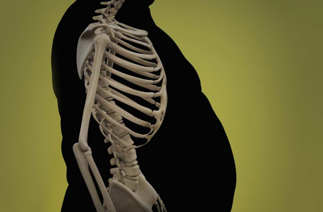
Visceral fat is body fat that is stored within the abdominal cavity and is therefore stored around a number of important internal organs such as the liver, pancreas and intestines.

Visceral fat is sometimes referred to as ‘active fat’ because research has shown that this type of fat plays a distinctive and potentially dangerous role affecting how our hormones function.

Storing higher amounts of visceral fat is associated with increased risks of a number of health problems including type 2 diabetes.

What types of fat do we store?

It is important to define the difference between visceral fat and subcutaneous fat. Subcutaneous fat is the fat that we store just under our skin. The fat we may be able to feel on our arms and legs is subcutaneous fat.

A growing belly can be the result of both types of fat. The fat we can feel just under the skin is subcutaneous fat but we may also be storing significant extra fat within our abdomen where our organs reside. This intra-abdominal fat is our visceral fat.

Visceral fat and insulin resistance

Carrying a high amount of visceral fat is known to be associated with insulin resistance, which can lead to glucose intolerance and type 2 diabetes. Researchers have found that visceral secretes a protein called retinol-binding protein 4 (RBP4) which has been shown to increase resistance to insulin.

What are the health risks associated with visceral fat?

All of us will have a certain amount of visceral fat but those of us with a larger quantity of visceral fat may be exposed to increased risks of the following health conditions.

  • Type 2 diabetes
  • Heart disease
  • Breast cancer
  • Colorectal cancer
  • Alzheimer’s disease

How can I tell if I have too much visceral fat?

The most certain way to tell if you are storing excess visceral fat is to undergo an MRI scan (Magnetic Resonance Imaging scan). However, MRI scans are not a cheap procedure and is therefore not recommended as a diagnosis tool for diabetes risk.

A relatively good indicator of visceral fat is to take a waistline measurement. Harvard University note around 10% of our total fat is likely to be stored as visceral fat, therefore if you are carrying higher amounts of body fat than is recommended, it is therefore more likely that you are also storing more visceral fat than is healthy.

Research has shown that the size of our belly is a relatively reliable indicator of the health risks linked to visceral fat.

Categories
blog

Reasons behind physical inactivity among diabetes patients

Researchers have found that fear of hypoglycemia (low blood sugar) act as major reason for physical inactivityamong diabetics. The study confirms earlier studies that found 60 percent of adult diabetics aren’t physically active.

Lack of understanding of insulin metabolism came alive when one hundred type 1 diabetes patients answered questionnaires to assess their barriers to physical activity. The biggest fear was hypoglycaemia and other barriers included interference with work schedule, loss of control over diabetes and low levels of fitness.

When questioned further, only 52 of the participants demonstrated appropriate knowledge of how insulin is metabolized and processed. Those individuals who best understood how insulin works in their body were shown to be less fearful of physical activity. Such knowledge is essential in order to adapt insulin and/or food intake to prevent hypoglycaemia induced by exercise.

Categories
blog

Hypertension, diabetes linked to carotid artery wall thickness


WASHINGTON – Researchers from A.O.U. in Cagliari Sardegna, Italy have found a link between increased carotid artery wall thickness (CAWT) – which can cause heart attack and stroke- and diabetes and hypertension.

During the study, the researchers looked at 186 patients with the help of multidetector row CT and sought to determine association between CAWT is associated with cardiovascular risk factors such as hypertension, diabetes mellitus, dyslipidemia and a history of smoking.

The results showed that there is a statistically significant relationship between diabetes and hypertension.

“There was no significant statistical correlation between the increase of carotid wall thickness, smoking and dyslipidemia,” said Dr Luca Saba, lead author of the study.

“Our group demonstrated that the presence of CAWT greater than 1mm in patients with diabetes or hypertension is strongly correlated with a risk to suffer a stroke. Patients at higher risk should be monitored every 12 months.

“The detection and evaluation of CAWT together with the study of other cardiovascular factors allows for improvement in patient follow up and risk stratification.

“Imaging as part of the diagnostic process and management of patients with high cardiovascular risks is important,” he added.

Categories
blog

Radiation therapy for childhood cancer may up diabetes risk

WASHINGTON – Childhood cancer survivors may have an increased risk of diabetes, particularly those who received total body or abdominal radiation, says a new study.

The study has been published in Archives of Internal Medicine, one of the JAMA/Archives journals.

To reach eth conclusion, Lillian R. Meacham, M.D., of Emory University and AFLAC Cancer Center and Blood Disorders Service, Atlanta, and colleagues compared the prevalence of diabetes in a sample of 8,599 childhood cancer survivors (diagnosed before age 21 between 1970 and 1986) and 2,936 randomly selected siblings of the survivors (average ages 31.5 and 33.4 at follow-up in 2003, respectively).

Medication use, treatment exposures (including irradiation, or exposure to radiation treatments) and factors that may have modified the risk of diabetes were noted.

Of the survivors, 218 (2.5 percent) reported having diabetes, while 49 (1.7 percent) of siblings reported having the condition.

“After adjustment for body mass index, age, sex, race/ethnicity, household income and insurance, the survivors were 1.8 times more likely than the siblings to report diabetes mellitus, with survivors who received total body irradiation, abdominal irradiation and cranial irradiation at increased risk,” the authors write.

“Survivors who were treated with abdominal irradiation were 2.7 times as likely to report diabetes mellitus as those who were not treated with abdominal irradiation or total body irradiation; those treated with total body irradiation were 7.2 times as likely to report diabetes mellitus,” they added.

Survivors diagnosed with cancer before age 5 were 2.4 times more likely to report diabetes than those diagnosed in late adolescence (from ages 15 to 20). (ANI)

Categories
blog

High-Fat Diet and Estrogen Loss Lead Women to Store More Abdominal Fat Than Men

A high-fat diet triggers chemical reactions in female mice that could explain why women are more likely than men to gain fat in the abdomen after eating excess saturated fat, new research suggests. The study also sheds light on why women gain fat following menopause.

Scientists identified events in female mice that start with the activation of an enzyme and end with the formation of visceral fat – fat that accumulates around internal organs and is linked to a higher risk for type 2 diabetes, heart disease and cancer.

At least one function for this enzyme is the production of a powerful hormone, which then drives up the formation of visceral fat cells. The source of this hormone is vitamin A.

This enzyme appears to be activated at higher levels in females than in males when both sexes eat a high-fat diet. When researchers genetically altered mice by deleting the enzyme, female mice stayed lean, especially in the abdominal area, even when they continued to eat a lot of fat. Males without the enzyme also developed less fat, but the effect was far less significant than in females.

The results suggest the enzyme could be a target for sex-specific anti-obesity therapy.

“If you asked most people what they believe causes obesity, they would probably say high food consumption and a sedentary lifestyle. But we see that there are genetic factors telling the body what to do with fat,” said Ouliana Ziouzenkova, assistant professor of human nutrition at Ohio State University and senior author of the study. “A high-fat diet acts on our genetics to make us more fat or less fat. The diet is not powerful enough to do this on its own.”

Further experiments showed that fat cells in female mice lacking the enzyme could produce proteins that use fat for heat, meaning the fat in females was burned away.

Researchers also studied fat tissue from human surgery patients and found the same enzyme was present in human tissue, and its levels were markedly higher in cells extracted from the visceral fat tissue of obese women compared to cells from lean women.

Finally, the study suggested that estrogen suppresses the enzyme’s activity, which might help explain why postmenopausal women with decreased estrogen in their bodies tend to accumulate fat in their bellies.

Categories
blog

Low HDL Cholesterol Linked to Diabetic Kidney Disease


Having a low level of HDL, or “good,” cholesterol, is a risk factor fordiabetic nephropathy (kidney disease)

According to new research published in the journal Diabetes Care. Diabetes is the leading cause of chronic kidney disease in the United States; between 5% and 15% of people with Type 2 diabetes, and between 25% and 40% of people with Type 1 diabetes, will someday develop diabetic nephropathy.

Diabetic nephropathy begins with changes to the glomeruli, or filtering units, of the kidneys. Over time, the tissue between the blood vessels of the glomeruli may expand, compressing the vessels and reducing their ability to filter waste products from the blood. Eventually, the glomeruli become a mass of scar tissue. Trace amounts of protein in the urine and high blood pressure are signs that a person may have nephropathy.

Low levels of HDL cholesterol are a known risk factor for atherosclerosis, or a buildup of fats and cholesterol in and on the artery walls, but there is little information about the link between low HDL and complications that involve the small blood vessels such as nephropathy. To evaluate the relationship between nephropathy,retinopathy(diabetic eye disease), and HDL cholesterol, researchers followed 11,140 people with Type 2 diabetes and at least one additional blood vessel–related risk factor for a median of five years as part of the Action in Diabetes and Vascular Disease: preterAx and diamicroN-MR Controlled Evaluation (ADVANCE) Study.

The researchers found that 3,585, or 32%, of the participants developed or experienced worsening of a microvascular condition over the follow-up period; 28% of the people had a kidney condition, while 6% had a retinal condition. The third of people with the lowest HDL cholesterol levels had a 19% higher risk of a kidney event than the third of people with the highest HDL cholesterol level. There was no link found between HDL cholesterol and retinopathy.

“Our findings provide the strongest evidence to date for a role of HDL-C [cholesterol] in the development and progression of diabetic nephropathy in patients with Type 2 diabetes,” the researchers note.

According to Harvard Medical School, strategies for raising your HDL levels include getting aerobic exercise such as swimming or bicycling, losing weight, stopping smoking, eating a healthful diet that does not include any trans fats, and taking a medicine such as niacin if recommended by your doctor